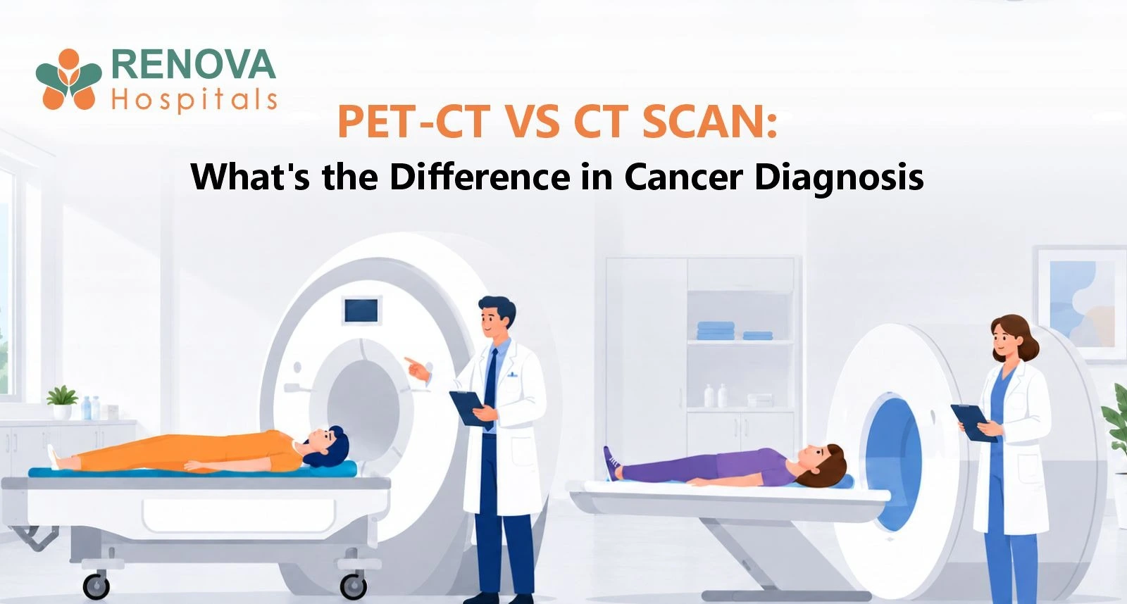
A CT scan primarily shows the structure, size, shape, and location of abnormalities inside the body. A PET-CT combines CT imaging with a PET scan, which shows areas of increased metabolic activity using a small amount of radioactive tracer. In cancer care, CT may help locate and measure tumours, while PET-CT may provide additional information about whether certain lesions are metabolically active and whether cancer has spread. Neither scan can independently confirm every cancer; biopsy and pathology are often required for definitive diagnosis.
Key Takeaways
- CT provides detailed anatomical images.
- PET shows metabolic activity within tissues.
- PET-CT combines metabolic and anatomical information.
- PET-CT is useful for selected cancers but is not necessary for every patient.
- Infection and inflammation can also appear active on PET.
- Not every cancer shows strongly on PET imaging.
- CT may be performed with or without contrast.
- Imaging can suggest cancer, but biopsy often establishes the definitive diagnosis.
What Is a CT Scan?
A
CT scan uses X-rays and computer processing to create detailed cross-sectional images of the body.
It can show:
- Organs
- Bones
- Blood vessels
- Lymph nodes
- Masses
- Areas of inflammation
In oncology, CT scans may help doctors:
- Identify suspicious masses
- Measure tumour size
- Assess nearby structures
- Look for spread to other organs
- Plan treatment
- Monitor response to therapy
CT is widely used in the evaluation of cancers affecting areas such as the chest, abdomen, and pelvis.
What Is a PET Scan?
For many cancer PET scans, a radioactive form of glucose known as FDG is used.
Because many cancer cells consume glucose at higher rates than surrounding tissues, they may accumulate more FDG and appear as areas of increased activity.
However, increased FDG uptake does not automatically mean cancer.
Inflammation and infection can also show increased uptake.
What Is a PET-CT Scan?
PET-CT combines two imaging technologies in a single examination.
PET Component
Provides information about metabolic activity.
CT Component
Shows the precise anatomical location of the abnormality.
Combining the two can help doctors determine where metabolically active tissue is located within the body.
What Is the Main Difference Between PET-CT and CT?
The simplest distinction is:
CT = structure
PET = activity
PET-CT = structure + metabolic activity
A CT scan may identify a lymph node that is enlarged.
PET imaging may provide information about whether that lymph node demonstrates increased metabolic activity.
However, neither finding should automatically be interpreted as
cancer without considering the clinical context.
When Is CT Used in Cancer Diagnosis?
CT may be used to:
- Investigate symptoms
- Characterise a suspicious mass
- Determine tumour location
- Assess lymph nodes
- Look for metastatic disease
- Guide certain biopsies
- Plan surgery or radiation treatment
- Monitor tumour size during treatment
When Is PET-CT Used in Cancer Care?
PET-CT may be useful for selected cancers when doctors need additional information about disease activity or distribution.
Possible uses include:
- Cancer staging
- Assessing suspected metastases
- Evaluating treatment response
- Investigating possible recurrence
- Helping distinguish active tumour from some post-treatment changes
- Planning certain radiation treatments
PET-CT is commonly used in selected patients with conditions such as:
Whether PET-CT is appropriate depends on the cancer type, stage, treatment history, and clinical question.
Is PET-CT Better Than CT for Cancer?
Not necessarily.
PET-CT and CT answer different questions.
A PET-CT is not simply a "better CT."
Some situations require:
- CT alone
- MRI
- PET-CT
- Ultrasound
- Mammography
- Endoscopy
- Bone scan
- Another specialised investigation
The best imaging test depends on the cancer being investigated.
Can PET-CT Detect All Cancers?
No.
Not all cancers show high FDG uptake.
PET sensitivity can vary according to:
- Cancer type
- Tumour size
- Tumour biology
- Blood glucose
- Location
- Previous treatment
Very small lesions may also be difficult to identify.
A normal PET-CT therefore does not rule out every cancer.
Can Infection Look Like Cancer on PET-CT?
Yes.
This is a critical limitation.
Inflammatory and infectious cells can also consume glucose and show increased FDG uptake.
Conditions such as infection, granulomatous inflammation, or recent procedures may therefore produce PET abnormalities.
A PET-positive lesion is not automatically cancer.
Further imaging, follow-up, or biopsy may be required.
Can CT Diagnose Cancer Without a Biopsy?
Imaging can strongly suggest cancer, but in many situations a definitive diagnosis requires tissue.
A
biopsy allows a pathologist to examine cells or tissue under a microscope and may provide information about:
- Cancer type
- Grade
- Molecular characteristics
There are exceptions where clinical management may differ, but patients should not assume that an imaging result alone always establishes the final diagnosis.
Does PET-CT Use Radiation?
Yes.
PET-CT involves radiation from:
- The radioactive tracer used for PET
- The CT component
CT also uses ionising radiation.
Doctors weigh the expected diagnostic benefit against radiation exposure when recommending imaging.
Why Is Blood Sugar Checked Before PET-CT?
FDG is related to glucose.
High blood glucose can affect how the tracer is distributed and may interfere with image quality.
Patients are commonly given specific instructions about:
- Fasting
- Diabetes medicines
- Insulin
- Exercise
- Appointment timing
Follow the imaging centre's instructions carefully.
People with diabetes should not independently change insulin or other medicines without appropriate guidance.
How Should You Prepare for PET-CT?
Instructions vary, but may include:
- Fasting for several hours
- Drinking plain water
- Avoiding strenuous exercise before the scan
- Informing staff about diabetes
- Informing staff about pregnancy or possible pregnancy
- Providing details of recent treatment or infection
After the tracer is injected, patients usually rest quietly before imaging.
How Long Does PET-CT Take?
The entire visit may take significantly longer than the actual scanning time because the tracer requires time to distribute through the body.
Timing varies between imaging centres and examination protocols.
Consult an Oncology Team at Renova Hospitals
Choosing the right cancer investigation depends on the suspected cancer, its location, previous test results, and the clinical question being answered.
At Renova Hospitals, our Medical Oncology, Surgical Oncology, Radiation Oncology, Nuclear Medicine, Radiology, and Pathology teams work together to determine the appropriate diagnostic and staging investigations for each patient.
Request a consultation or call 040 2333 3333.
When Is Prompt Medical Evaluation Important?
Seek medical evaluation for persistent or unexplained symptoms such as:
- Unexplained weight loss
- A growing lump
- Persistent unusual bleeding
- Blood in stool or urine
- Persistent cough
- Difficulty swallowing
- Unexplained lymph-node enlargement
- Symptoms that progressively worsen
Imaging should be selected based on clinical evaluation rather than requesting PET-CT as a general cancer check.
Medical References
Medically Reviewed & Approved By:
Dr. G. Reshma Reddy
MBBS, MD (Radio-Diagnosis), Fellowship in Newer Imaging – Senior Consultant Radiologist
This article has been reviewed and approved by a qualified medical professional to ensure medical accuracy and adherence to current clinical standards.