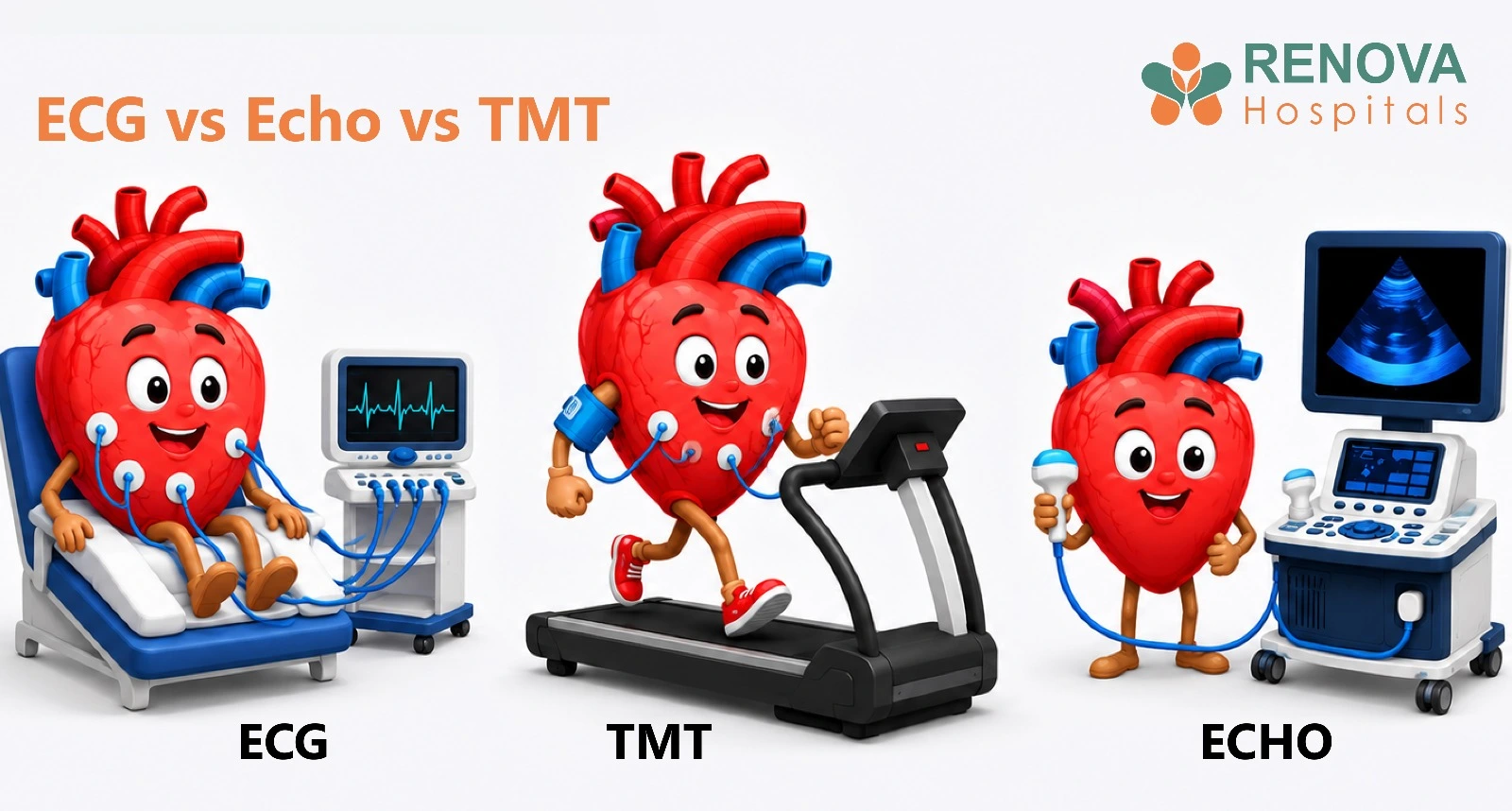
An ECG records the heart’s electrical activity and rhythm. An echocardiogram, commonly called an Echo or 2D Echo, uses ultrasound to examine the heart’s chambers, valves and pumping function. A TMT evaluates how the heart responds during exercise. These tests answer different clinical questions, and a
cardiologist may recommend one or more based on your symptoms, examination findings and cardiovascular risk.
Key Takeaways
- ECG checks the heart’s rate, rhythm and electrical signals.
- Echo or 2D Echo examines the heart’s structure, valves, muscle movement and pumping function.
- TMT assesses how the heart responds while you exercise under medical supervision.
- A TMT may suggest reduced blood supply during exertion, but it does not directly show or confirm a coronary artery blockage.
- A normal result on one test does not rule out every heart condition.
Seek emergency medical care
New or severe chest pressure, chest pain at rest, sudden breathlessness, sweating, fainting, weakness, difficulty speaking, confusion, or pain spreading to the arm, back, neck or jaw may indicate a medical emergency. Do not wait for a routine ECG, Echo or TMT appointment.
What Is the Main Difference Between ECG, Echo and TMT?
The simplest way to understand the difference is:
- ECG checks how the heart’s electrical system is working.
- Echo checks how the heart looks and how effectively it pumps.
- TMT checks how the heart responds when physical effort increases.
These tests are complementary. They are not interchangeable, and one is not automatically better than the others.
What Is an ECG?
An electrocardiogram, also called an ECG or EKG, records the electrical signals produced during each heartbeat. Small electrodes are placed on the chest and sometimes on the arms and legs. The recording shows how fast the heart is beating, whether the rhythm is regular and how electrical signals travel through different parts of the heart.
What Can an ECG Help Identify?
- A fast heart rate, known as tachycardia
- A slow heart rate, known as bradycardia
- Irregular heart rhythms or arrhythmias
- Electrical conduction abnormalities
- Certain patterns associated with heart strain or heart-muscle injury
- Signs that may be associated with a current or previous heart attack
- Changes related to some electrolyte disturbances or medicines
- The functioning of certain implanted cardiac devices
An ECG is often part of the initial evaluation when a patient experiences chest discomfort, palpitations, dizziness, fainting or unexplained breathlessness.
What Can an ECG Not Show?
- It does not create an image of the heart.
- It does not directly show the heart chambers or valves.
- It does not directly visualise coronary artery blockages.
- A short resting ECG may miss an irregular rhythm that occurs only occasionally.
- A normal ECG does not rule out every heart condition.
When symptoms such as palpitations occur intermittently, a cardiologist may recommend longer rhythm monitoring, such as a Holter monitor or event monitor.
How Should You Prepare for an ECG?
- Wear clothing that allows easy access to the chest.
- Avoid applying creams or oils to the chest before the test.
- Inform the medical team about your current medicines and implanted cardiac devices.
- Remain still while the recording is taken.
An ECG is non-invasive and usually painless.
What Is an Echocardiogram?
An echocardiogram, commonly called an Echo or 2D Echo, is an ultrasound examination of the heart. Gel is applied to the chest and a device called a transducer is moved over different areas. Sound waves are converted into moving images that show the heart’s structure, muscle movement, valves and blood flow.
What Can an Echo Help Evaluate?
- The size of the heart chambers
- The thickness and movement of the heart muscle
- The heart’s pumping ability and ejection fraction
- Narrowed or leaking heart valves
- Abnormal movement of parts of the heart muscle
- Fluid in the sac surrounding the heart
- Certain blood clots within the heart chambers
- Some congenital heart abnormalities
- Changes associated with heart failure or cardiomyopathy
What Is Ejection Fraction?
Ejection fraction estimates how much blood the left ventricle pumps out with each contraction. It is one of several measurements used to assess pumping function. The result must be interpreted with other findings because a single number does not provide a complete picture of heart health.
What Can an Echo Not Show?
- A standard resting Echo does not directly show most coronary artery blockages.
- It does not continuously record intermittent rhythm problems.
- It cannot identify every possible cause of chest discomfort.
- Image quality may be limited in some patients because of body structure, lung disease or other technical factors.
- A normal Echo does not rule out every heart condition.
When May a Doctor Recommend an Echo?
- Unexplained breathlessness
- A heart murmur or suspected valve disease
- Swelling in the legs with suspected cardiac involvement
- Heart failure or cardiomyopathy
- A previous heart attack
- Abnormal findings during a cardiac examination
- Certain abnormal ECG findings
- Monitoring an existing heart condition
How Should You Prepare for an Echo?
A standard transthoracic Echo usually requires little or no special preparation. Patients can generally eat, drink and take their usual medicines unless instructed otherwise. Other types, including transoesophageal Echo or stress Echo, may require fasting or additional preparation.
What Is a TMT?
A treadmill test, also called a TMT, exercise ECG or exercise stress test, assesses how the heart responds as physical effort gradually increases. Electrodes monitor the ECG while a blood-pressure cuff records readings. The patient walks on a treadmill as its speed and incline increase in stages.
During the test, the medical team monitors:
- Heart rate and rhythm
- Blood-pressure response
- Breathing and exercise tolerance
- Chest discomfort, dizziness or other symptoms
- ECG changes during exercise and recovery
What Can a TMT Help Evaluate?
- Chest discomfort that appears during exertion
- Unexplained breathlessness during physical activity
- Exercise tolerance
- Heart-rate and blood-pressure response to exercise
- Exercise-triggered rhythm disturbances
- Possible exercise-induced myocardial ischaemia
- Response to selected cardiac treatments
- Safe exercise capacity in selected patients
Can a TMT Detect a Heart Blockage?
A TMT does not directly show a coronary artery blockage. It may identify symptoms or ECG changes during exercise that suggest reduced blood supply to the heart muscle. An abnormal result does not automatically confirm whether a blockage is present, where it is located or how severe it is.
Depending on the patient’s symptoms and overall risk, a cardiologist may consider additional investigations such as:
- Stress echocardiography
- Myocardial perfusion imaging
- Coronary CT angiography
- Invasive coronary angiography
The next investigation is chosen after reviewing the complete clinical picture rather than the TMT result alone.
Who May Not Be Suitable for a TMT?
A TMT is not suitable for every patient. A cardiologist will assess whether exercising on a treadmill is safe and whether the test is likely to provide useful information. It may not be appropriate for some people with:
- An acute or unstable heart condition
- Uncontrolled high blood pressure
- Certain serious rhythm abnormalities
- Severe symptomatic valve disease
- Significant difficulty walking
- Severe joint, neurological or respiratory limitations
- Other conditions that make exercise unsafe
Alternative forms of stress testing may be considered when a patient cannot exercise adequately.
How Should You Prepare for a TMT?
- Wear comfortable clothing and suitable walking shoes.
- Avoid a heavy meal immediately before the test.
- Ask your doctor whether you should take your regular medicines.
- Do not stop heart or blood-pressure medicines without medical advice.
- Inform the team about chest pain, breathing difficulty, joint problems or mobility limitations.
- Avoid strenuous exercise shortly before the test unless instructed otherwise.
The test may be stopped if concerning symptoms, significant ECG changes, an abnormal blood-pressure response or severe fatigue develops.
Which Heart Test May Be Considered for Different Symptoms?
Palpitations or an Irregular Heartbeat
An ECG may be part of the initial assessment because it records the heart’s rhythm. Longer rhythm monitoring may be needed when symptoms occur only occasionally.
Breathlessness
An Echo may help assess pumping function, valve function and heart-muscle movement. Other tests may be required because breathlessness can have cardiac and non-cardiac causes.
A Heart Murmur
An Echo may be recommended to examine the valves and determine whether there is narrowing, leakage or another structural abnormality.
Chest Discomfort During Exercise
A TMT or another form of stress testing may be considered after clinical evaluation. The most appropriate test depends on age, symptoms, exercise capacity, ECG findings and overall cardiovascular risk.
Chest Pain at Rest
Chest pain at rest requires urgent medical evaluation, especially when it is new, severe or accompanied by sweating, breathlessness, fainting or weakness. Assessment may include an ECG, blood tests such as troponin and other investigations.
Unexplained Fainting
An ECG may form part of the initial evaluation. Additional rhythm monitoring, imaging or other investigations may be required based on the suspected cause.
Symptoms alone cannot determine which test is appropriate. A clinical evaluation is necessary.
Is an Echo Needed After a Normal ECG?
Not always. A normal ECG does not automatically mean that an Echo is required or unnecessary. An Echo may be considered when symptoms, a heart murmur, examination findings or the patient’s history suggest a possible structural or pumping problem. The tests answer different questions, so the decision is based on clinical need rather than the ECG result alone.
Why May a Cardiologist Order More Than One Test?
A cardiologist may recommend more than one investigation because each test provides different information:
- An ECG may assess rhythm and electrical signals.
- An Echo may examine heart muscle, valves and pumping function.
- A TMT may assess symptoms and ECG changes during exercise.
- Blood tests or advanced imaging may be required when the initial findings do not provide a complete answer.
The sequence depends on symptoms, age, medical history, physical examination, cardiovascular risk, ability to exercise and initial test results.
Important Limitations of ECG, Echo and TMT
ECG Limitations
- A short resting ECG may miss intermittent rhythm abnormalities.
- It does not directly show heart structure or coronary artery blockages.
- A normal result does not rule out every heart problem.
Echo Limitations
- It does not directly show most coronary artery blockages.
- It does not continuously monitor intermittent rhythm abnormalities.
- Image quality may vary between patients.
TMT Limitations
- It may produce false-positive or false-negative findings.
- It does not directly visualise the coronary arteries.
- Its usefulness may be limited when the patient cannot exercise adequately.
- It is not suitable for every patient.
All test results must be interpreted alongside symptoms, clinical examination and medical history.
Myth vs Fact
Myth: A Normal ECG Means the Heart Is Completely Healthy
Fact: A normal ECG does not rule out every heart condition. Further evaluation may be needed when symptoms continue or the clinical risk remains significant.
Myth: A TMT Directly Shows Blocked Arteries
Fact: A TMT may suggest reduced blood supply during exercise, but it does not directly visualise or confirm a coronary artery blockage.
Myth: ECG and Echo Are the Same Test
Fact: An ECG records electrical activity, while an Echo uses ultrasound to examine structure and pumping function.
Myth: Everyone With Diabetes or High Blood Pressure Needs All Three Tests
Fact: Cardiovascular risk factors do not automatically mean that every heart test is required. Testing should be individualised. Routine resting or exercise ECG screening is not recommended for asymptomatic adults at low cardiovascular risk.
Conclusion
ECG, Echo and TMT evaluate different aspects of heart health. An ECG records electrical activity, an Echo examines structure and pumping function, and a TMT assesses how the heart responds during exercise. None is automatically better than the others, and no single test can diagnose every heart condition. The appropriate investigation depends on your symptoms, medical history, examination findings and overall cardiovascular risk.
Consult a cardiologist if you experience chest discomfort, breathlessness, palpitations, fainting or an unexplained reduction in exercise capacity. Seek emergency medical care for new or severe chest pain, especially when it occurs at rest or is accompanied by sweating, breathlessness, fainting, weakness or difficulty speaking.
Consult a Cardiologist at Renova Hospitals
Renova Hospitals provides comprehensive cardiac evaluation and personalised guidance for ECG, Echo, TMT and other heart investigations.
For cardiology appointments and enquiries, call 040 2333 3333
Medical References
Medically Reviewed & Approved By:
Dr. B.K. Srinivasa Sastry
Degrees : MBBS, MD (Internal Medicine), DM (Cardiology)
Experience : 40 years of experience as interventional cardiologist
This article has been reviewed and approved by a qualified medical professional to ensure accuracy, credibility, and adherence to current clinical standards.